
Պատկեր:Computed tomography of human brain - large.png

Սկզբնական նիշք (3639 × 2595 կէտիկներ, նիշքի չափը՝ 3,9 ՄԲ, MIME-տեսակը՝ image/png)
| This file is made available under the Creative Commons CC0 1.0 Universal Public Domain Dedication. | |
| The person who associated a work with this deed has dedicated the work to the public domain by waiving all of their rights to the work worldwide under copyright law, including all related and neighboring rights, to the extent allowed by law. You can copy, modify, distribute and perform the work, even for commercial purposes, all without asking permission.
|
|

| Նկարագրութիւն |
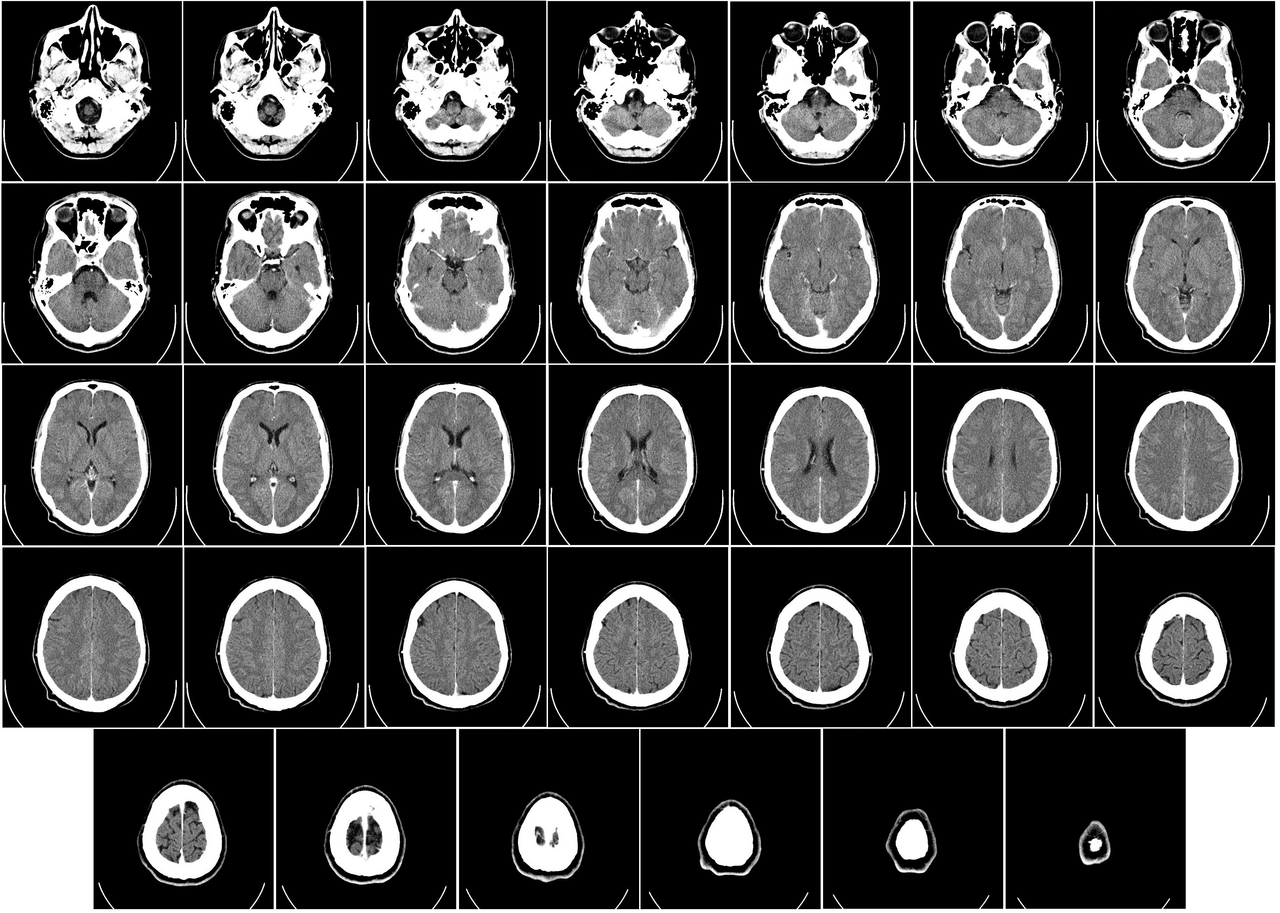
English: Computer tomography of human brain, from base of the skull to top. Taken with intravenous contrast medium.
It was taken Mars 23, 2007 on the uploader, after a 20 minute episode of homonymous hemianopsia with loss of the left visual field, but nothing strange was found. Three episodes of scotoma occurred in the following years, whereof the last one was scintillating (depiction). Otherwise, there were no further neurological symptoms.
Türkçe: Geçirdiği bir kaza neticesinde homonim hemianopsi vakası oluşan bir hastanın beyninin bilgisayarlı tomografisi. Tomografi neticesinde bir anomaliye rastlanmamıştır. |
| Թուական | Uploaded January 17, 2008 |
| Աղբիւր | Radiology, Uppsala University Hospital. Uploaded by Mikael Häggström. |
| Հեղինակ | Department of Radiology, Uppsala University Hospital. Uploaded by Mikael Häggström. |
| Արտօնութիւն (Նիշքի վերօգտագործում) |
Compound images
-

-
 Inverted


Scrollable stack
For larger version, see Category:Computed tomography images of Mikael Häggström's brain. To move through the images, hover over the image and use scroll wheel, drag the mouse, or click the < or the > above each stack. This functionality should activate when the page is fully loaded, which may take some time.

.png)
.png)
.png)
.png)
.png)
.png)
.png)
.png)
.png)
.png)
.png)
.png)
.png)
.png)
.png)
.png)
.png)
.png)
.png)
.png)
.png)
.png)
.png)
.png)
.png)
.png)
.png)
.png)
.png)
.png)
.png)
.png)
.png)
.png)
Case with multiplanar reconstruction
-
 Brain, case 1: Multiplanar, but no intravenous contrast.
Individual images
Licencing
| This file is made available under the Creative Commons CC0 1.0 Universal Public Domain Dedication. | |
| The person who associated a work with this deed has dedicated the work to the public domain by waiving all of their rights to the work worldwide under copyright law, including all related and neighboring rights, to the extent allowed by law. You can copy, modify, distribute and perform the work, even for commercial purposes, all without asking permission.
|
DICOM format
Նիշքի պատմութիւն
Սեղմել օրուան/ժամին վրայ նիշքի այդ պահուն ունեցած վիճակը տեսնելու համար
| Օր/Ժամ | Մանրապատկեր | Ծաւալ | Գործածող | Մեկնաբանութիւն | |
|---|---|---|---|---|---|
| ընթացիկ | 01:11, 24 Դեկտեմբեր 2017 | | 3639 × 2595 (3,9 ՄԲ) | Shashi. | Reverted to version as of 12:49, 1 February 2008 (UTC) |
| 10:59, 8 Մայիս 2008 |  | 3639 × 2595 (3,17 ՄԲ) | CountingPine | Optimise using PNGOUT | |
| 12:49, 1 Փետրուար 2008 |  | 3639 × 2595 (3,9 ՄԲ) | Mikael Häggström | {{34 computer tomography images}} {{Individual images of CT of Mikael Häggström's brain}} | |
| 11:56, 31 Յունուար 2008 |  | 3639 × 2595 (4,03 ՄԲ) | Mikael Häggström | {{34 computer tomography images}} {{Individual images of CT of Mikael Häggström's brain}} |
Նիշքի գործածութիւն
Հետեւեալ էջը կը յղուի այս նիշքին՝
Նիշքի համընդհանուր օգտագործում
Հետեւեալ ուիքիները եւս կ'օգտագործեն այս նիշքը՝
- Օգտագործումը bn.wikipedia.org կայքին վրայ
- Օգտագործումը bo.wikipedia.org կայքին վրայ
- Օգտագործումը ca.wikipedia.org կայքին վրայ
- Օգտագործումը en.wikipedia.org կայքին վրայ
- CT scan
- Portal:Medicine
- Portal:Medicine/Selected picture
- Portal:Medicine/Selected picture archive
- Wikipedia:WikiProject Neuroscience
- Wikipedia:Featured pictures/Sciences/Biology
- User:Mikael Häggström
- User talk:Mikael Häggström/Archive 1
- Wikipedia:Featured pictures thumbs/10
- Wikipedia:Featured picture candidates/CT of brain of Mikael Häggström.png
- Wikipedia:Featured picture candidates/February-2008
- Wikipedia:Wikipedia Signpost/2008-02-11/Features and admins
- Portal:Medicine/Selected picture/9, 2008
- Portal:Medicine/Selected picture/9
- Wikipedia:Picture of the day/July 2008
- Template:POTD/2008-07-11
- Wikipedia:Wikipedia Signpost/2008-02-11/SPV
- User:Mikael Häggström/Gallery
- Wikipedia:WikiProject Medicine/Recognized content
- Computed tomography of the head
- Wikipedia:Wikipedia Signpost/2013-10-02/Op-ed
- Wikipedia:Wikipedia Signpost/Single/2013-10-02
- User:Wouterstomp/test
- User:Fitness queen04/sandbox
- Wikipedia:WikiProject Anatomy/Resources
- Wikipedia:WikiProject Anatomy/Recognized content
- Wikipedia talk:WikiProject Anatomy/Archive 9
- Reconstruction from projections
- User:VGrigas (WMF)/Quality Media
- User:Flyer22 Frozen/Human brain
- Portal:Medicine/Recognized content
- User talk:Rhododendrites/Reconsidering FPC on the English Wikipedia
- Օգտագործումը es.wikipedia.org կայքին վրայ
- Օգտագործումը fi.wikipedia.org կայքին վրայ
- Օգտագործումը he.wikipedia.org կայքին վրայ
- Օգտագործումը hy.wikipedia.org կայքին վրայ
- Օգտագործումը id.wikipedia.org կայքին վրայ
- Օգտագործումը is.wikipedia.org կայքին վրայ
- Օգտագործումը ja.wikipedia.org կայքին վրայ
- Օգտագործումը ka.wikipedia.org կայքին վրայ
Տեսնել այս նիշքի աւելի համընդհանուր օգտագործումը: